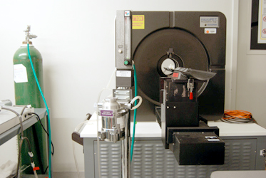
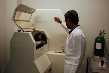
CIR has available microPET and microCT modalities for use by investigators for in-vivo/ex-vivo studies. Full-service is available for all modalities, where our technicians perform the imaging and/or processing and provide the data to you. Training is also available for post-processing services. PET radiotracer and radiolabeling services supported by our in-house cyclotrons are also available in our center.
CIR faculty and technicians are available for technical support in: Study design, Imaging protocols and techniques, Post-processing and data analysis, Data interpretation, and Training.
Positron Emission Tomography (PET) Imaging
PET Imaging is essential for molecular imaging research. It is a nuclear medicine functional imaging technique that is used to observe metabolic processes in the body. The PET scanner uses a ring of highly sensitive crystals to detect pairs of gamma rays emitted by a positron-emitting radionuclide (tracer), which is introduced into the body on a biologically active molecule. The scanner produces a 3 dimensional image of the anatomic distribution of the radioactive tracer and can measure the rates of biochemical processes in-vivo using tracer kinetics.
Services Available:
- Training of faculty, students, and personnel on imaging software and analysis.
- Research applications for microPET imaging of small animals
Computed Tomography (CT) Imaging
Computed tomography (CT), formerly referred to as computed axial tomography (CAT), uses X-rays to produce detailed cross-sectional images of the body. Multiple X-ray projections are reconstructed using a filtered back projection reconstruction technique into a 3D image of the object in the scanner. Clinical CT imaging is used to produce detailed anatomic images of the body and is particularly useful for imaging of the bone. It is routinely used clinically in PET/CT scanners to produce low-dose CT images used to calculate attenuation correction and then fused with the PET images to show detail in anatomic structures. In the same way, our microCT scanner images can be fused with our microPET images to provide enhanced anatomical detail.
Services Available:
- Training of faculty, students, and personnel on imaging software and analysis.
- Research applications for CT imaging of small animals or objects


