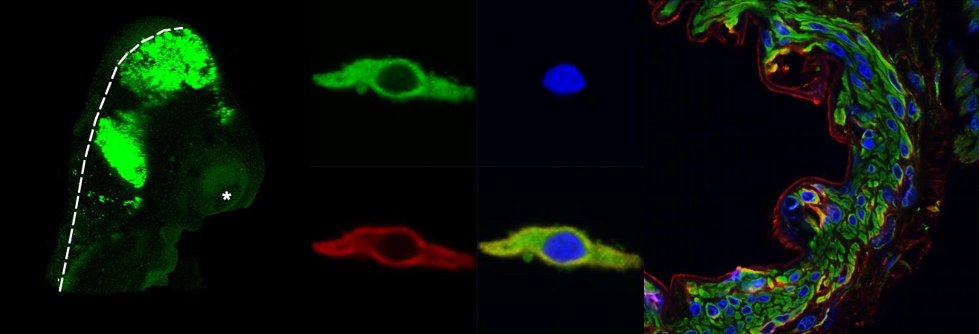
Recent advances in light microscopy techniques and associated technologies have allowed researchers in varied disciplines to manipulate and derive information to advance our understanding of the mechanisms associated with cell and tissue structure and function. Traditional light microscopy coupled with fluorescence imaging has enabled researchers to quantify their samples and understand basic biological processes. Laser scanning confocal microscopy coupled with multispectral analysis and multiphoton capabilities provides even more precise sample visualization and exceptional information extraction.
The Advanced Imaging and Microscopy Core Facility has four microscopy systems. There are two inverted laser scanning confocals, a stereologer system, an epifluorescence/brightfield system. In addition, there are 3 analysis computer workstations where users can analyze their data. The Imaris software on one of the workstations has allowed scientists around the world with publishing cutting edge research in high impact journals. The software is great for altering and analyzing fluorescence 3D images. The Visiopharm software, found on another one of the workstations is particularly helpful in analyzing 2D brightfield images. It has specific APPs which can be modified or reinvented depending on what type of analysis is needed.
What services do we offer?
- Training of faculty, students, and personnel on various microscope systems and image analysis software.
- Full service imaging option available.
- Research applications for image acquisition and photo microscopy.
- Research applications for Confocal LSM image analysis with Zeiss Zen software
- Other image analysis capabilities with MediaCybernetics Image Pro Plus 6.3, NIH Image, Imaris and Visiopharm.
- Referral for sample preparation, staining, and immunohistochemistry processing.
