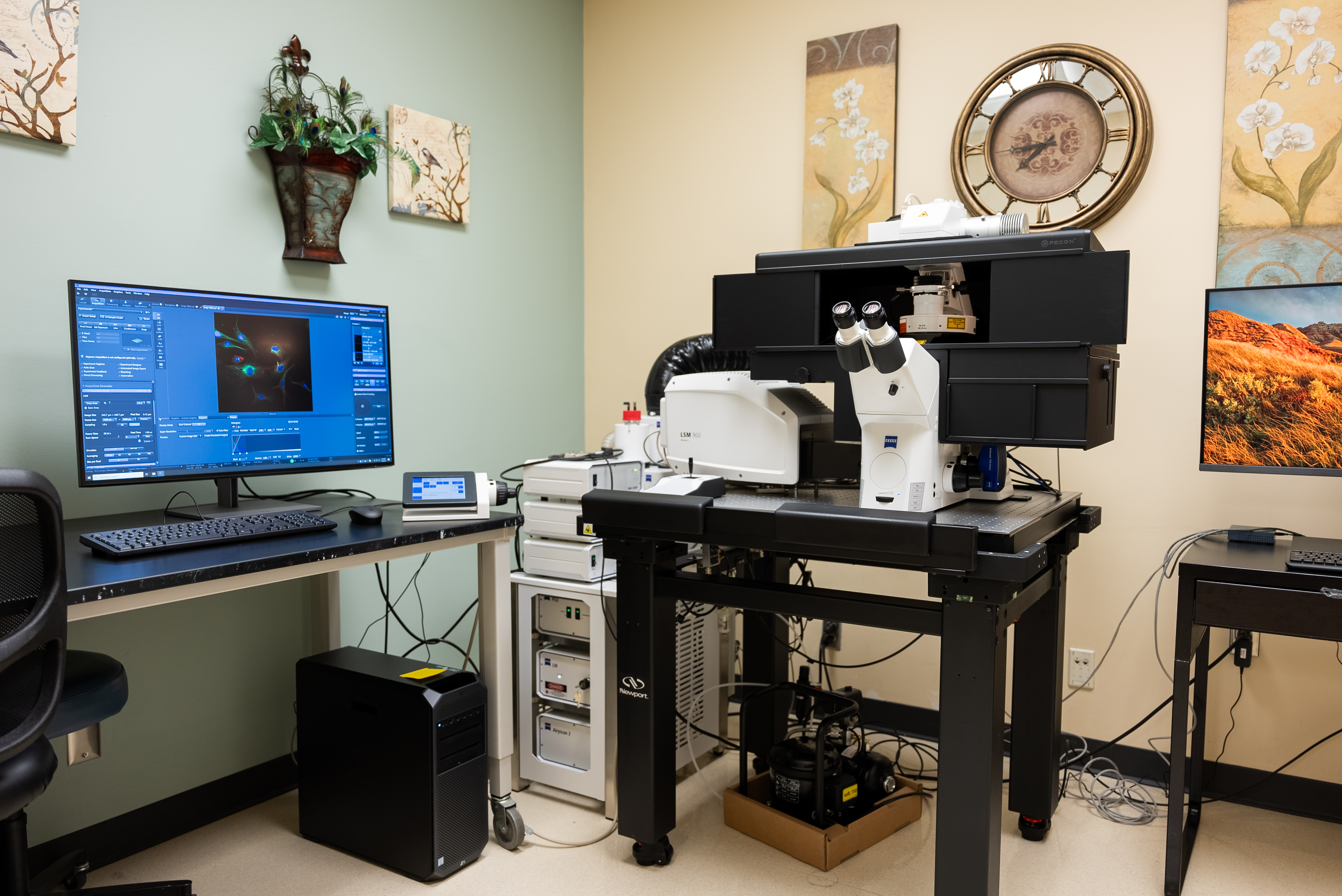
This inverted (Axio Observer 7) confocal imaging workstation produces super-resolution, high-contrast images while minimizing light exposure, to reduce photobleaching and channel bleed-through. The system has a diode laser module allowing for light emission at 405 nm, 488 nm, 561 nm, and 640 nm. It utilizes two GaAsP PMT detectors, one transmitted light detector, and an Airyscan 2 detector with Multiplex Mode for super-resolution imaging along with adaptive focusing capabilities. The Airyscan 2 is an area detector with 32 detection elements which allows for rapid confocal imaging for spatial and temporal recordings, as well as generating mosaic tiling for multidimensional reconstruction. The microscope performs deconvolution using LSM Plus and Joint Deconvolution (jDCV), using a dedicated computer workstation. LSM Plus uses a Wiener filter to enhance signal noise, improve resolution, and provide spatial information. jDCV works alongside the Airyscan 2 detector to add additional structural information and further improve resolution.
The system uses an Axiocam 705 mono, which provides high-resolution monochrome image capture, low light fluorescence imaging, high frame rate imaging, and broad spectral sensitivity from UV to near-IR light. The microscope is equipped with a motorized stage suitable for tiling of large areas and a Z piezo stage to generate Z-stacks with precision. For multi-day, multi-position time-lapses, the system utilizes Definite Focus 3 to compensate for focus drift, resulting in sharp, high-contrast images. An opaque, integrated incubation module creates stable temperature conditions when imaging temperature-sensitive samples and eliminates the effects of potential ambient light. The objectives include EC-Plan NEO 2.5X 0.085 NA; Plan-APO 10X 0.45 NA; Plan-APO 20X 0.8 NA; Plan-APO 40X 0.95 NA; C-APO 40X 1.2 NA W; Plan-APO 63X 1.4 NA Oil. The purchase of the Zeiss 900 LSM for the Advanced Imaging and Microscopy Core Facility in the School of Medicine was made possible due to the support of NIH S10OD034427.
This system is ideal for observing cell signaling, in-situ hybridization, protein expression and localization, interactions between molecules and proteins, spatial reconstruction, spatial-temporal relationships, and subcellular transport.
News Article: Revolutionizing Research: School of Medicine Unveils Cutting-Edge Super-Resolution Microscope at Advanced Imaging and Microscopy (AIM) Core Facility.
